


to Friday, August 8, 2025
Conference Registration
We use email to track your registration, so make sure to use the same email address every time!
A Message from Dr. Betty Kutter
Greetings, my dear phage family,
More than 50 years ago, we built the Evergreen Phage Meeting as a place for discovery, collaboration, and the relentless pursuit of phage science. What a journey it has been! Though I won’t be there in person this year, my heart is with you all as you continue to push the field forward.
Evergreen has been one of the greatest experiences of my life, and I am grateful to see the Phagebiotics Research Foundation carrying this legacy into the future. Keep asking big questions, forging new connections, and sharing knowledge—because that’s what makes this community truly special.
Wishing you all a fantastic meeting and many more to come!
With gratitude,
Dr. Betty Kutter
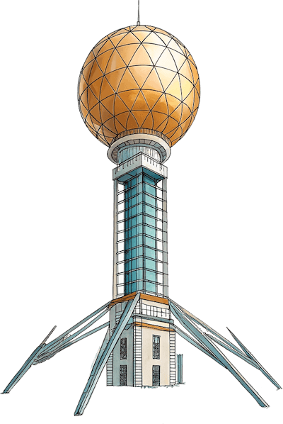
Evergreen Phage Meeting heads to Knoxville
After 50 years in Olympia, WA, the Evergreen Phage Meeting is hitting the road for the first time with its first stop in Knoxville, Tennessee! Abstracts for both oral and poster presentations can be submitted up until July 7th but will be accepted on a rolling basis.
📅 August 3-8, 2025 | Knoxville, TN
Organized by Phagebiotics Research Foundation
Led by Ria Kaelin (Phagebiotics) & Tom Denes (UT Knoxville) in collaboration with PhageDirectory
A meeting for all phage researchers
The Evergreen Phage Meeting welcomes researchers at all career stages, from students to seasoned scientists. We invite participants from around the world and value contributions from a wide range of research environments, recognizing the importance of diverse perspectives and resource conditions in advancing phage science.
🎟 Your All-Access Registration Includes
- Workshops (Sunday)
- Full Scientific Program (Mon - Thur)
- Breakfast Grab Bag -(Mon–Thur) Breakfast option for your hotel stay
- Catered Lunch at UT Gardens (Sun–Thurs) + Coffee, Tea & Snack
- Legacy Gala Dinner held in downtown Knoxville (Wednesday Night)
Join us for a memorable evening at The Press Room as we celebrate the pioneers of phage biology, with a special tribute to Dr. Betty Kutter for her contributions to the Evergreen Phage Community.
- Two Poster Sessions – (Tue & Thur) Apps, Wine & Cheese + Ice Cream Social
- Film Screening — Last Chance to Save a Life (IMDB) + Panel Discussion | August 4 | Bijou Theater (est. 1909)
- Digital Abstract Book
- Hotel-to-Conference Transportation
- Evergreen 2025 T-shirt & Sticker
Friday social add-on (Aug 8)
Trip to Gatlinberg and Anakeesta:
We will provide transport to the mountain town of Gatlinberg and admission to the mountaintop park Anakeesta .
Program
Some of the sessions and dates may change as we adjust to the volume of submitted abstracts. We will try to keep the final program close to this schedule
Sunday (08/03) - Workshops
Phage Genomics in the AI era: Ramy K. Aziz, Professor of Microbiology & Immunology, Faculty of Pharmacy, Cairo University and Head of MARC Biolabs, Egypt
Overview of Phage Purification Techniques: Tobi Nagel, Founder and President, Phages for Global Health
Viromics workshop: Evelien Adriaenssens, Group Leader, Quadram Institute of Bioscience, Norwich, Great Britain
This workshop is for participants who want to learn the principles of viromics (viral metagenomics) bioinformatics analyses, particularly focused on the bacteriophage complement of the microbiome. The workshop will consist of a mixture of lecture and hands-on analyses. Hands-on analyses will be performed using a command-line interface and free software on a publicly available dataset. Experience in operating a command-line environment is ideal but the workshop will be open to interested participants from all backgrounds and levels of expertise.
Evening: Informal meetup in downtown Knoxville, TBD
Monday (08/04)
- Introduction
- Plenary Talk: Steven Wilhelm, Kenneth & Blaire Mossman Professor, Department of Microbiology, University of Tennessee
- Catered lunch at the UT Gardens
- Session: Ecology and Evolution
- Evening Event: Screening of "Last Chance to Save a Life" at the Bijou Theater
Tuesday (08/05)
- Plenary Talk: Joseph Bondy-Denomy, Associate Professor, Microbiology and Immunology, University of California, San Francisco
- Session: Phage-Host Interactions
- Catered Lunch at the UT Gardens
- Session: Proteomics, Genomics, and Molecular mechanisms
- Poster Session with Wine and Cheese
Wednesday (08/06)
- Session: Phage Medical Applications - 1
- Catered Lunch at the UT Gardens
- Session: Phage Medical Applications - 2
- Session: Industry Speakers and Roundtable
- Evening Event: Gala Dinner at The Press Room
Thursday (08/07)
- Session: Agriculture and Food Safety
- Catered Lunch at the UT Gardens
- Poster Session with Ice Cream
- Session: Closing Matters
Friday (08/08) - Optional
Day-trip to Gatlinberg and Anakeesta mountain park in the Great Smoky Mountains. A more strenuous hiking option will be available by inquiry for the more adventurous.
Confirmed Invited Speakers (list only includes invited speakers)
- Plenary Talk: Steven Wilhelm, Kenneth & Blaire Mossman Professor, Microbiology, University of Tennessee
- Plenary Talk: Joseph Bondy-Denomy, Associate Professor, Microbiology and Immunology, University of California, San Francisco
- Joanne Emerson, Plant Pathology, University of California, Davis
- Alison Buchan, Microbiology, University of Tennessee, Knoxville
- James Gurney, Biology, Georgia State University
- Bryan Hsu, Biological Sciences, Virginia Tech
- Katrine Whiteson, Dept Mol Biol & Biochem, University of California, Irvine
- Evelien Adriaennsens, Quadram Institute of Bioscience
- Patrick Needham, Food Science, Cornell University
- Terje Dokland, Microbiology, University of Alabama
- Mark Radosevich, Biosystems Engineering and Soil Science, University of Tennessee
- Steven Bowden, Food Science, University of Minnesota
- Hany Anany, Agriculture and Agri-Food Canada
Join us as we launch a new chapter
This marks a major transition for the Evergreen Phage Meeting. Your participation and contribution help ensure the meetings success for years to come 🚀 Join us in Knoxville.
Questions? Contact us—we’re here to help. tdenes@utk.edu
Abstracts (due July 7th)
Accepted abstracts will be assigned to one of the following sections:
- Phage Ecology and Evolution
- Agriculture and Food Safety
- Phage-host Interactions
- Proteomics, Genomics and Molecular mechanisms
- Phage Medical Applications
- Industry
Please submit your abstract as soon as possible, as they are accepted on a rolling basis.
If you need a Visa Letter, you must submit your abstract before requesting a visa letter!
All abstracts have a 4,000 character limit for the body of the abstract.
Abstract Deadlines:
- Oral Presentation abstracts are due JULY 7th 2025
- Poster abstracts are due JULY 7th 2025
Posters and Printing
- Poster sizes: up to 36 (width) ×48 inches (height) and can be printed in Knoxville for $72. Matte paper type with no lamination and no mounting is preferred. Upload your posters to be printed here: https://local.fedex.com/en-us/tn/knoxville/office-0501 by July 31, and we will pick them up and have them ready for you by check-in.
- Double check that your selected address is: 2010 Cumberland Ave. Suite 4 Knoxville, TN 37916 Please indicate for in-store pickup: Phagebiotics or Dan Bryan
Pricing
For groups of 3 or more, email Ria at ria@phagebiotics.org for a group discount!
| Early Bird Academic & Government | $750 USD |
| Early Bird Industry | $1,000 USD |
| Student | $750 USD |
| Academic & Government | $850 USD |
| Industry | $1100 USD |
Hotel Pricing
Book your Hotel by JULY 12th to have booking rate below.
Marriott Discount link: https://www.marriott.com/event-reservations/reservation-link.mi?id=1741788650055&key=GRP&guestreslink2=true&app=resvlink
Hilton Discount link: https://www.hilton.com/en/book/reservation/rooms/?ctyhocn=KNXKHHF&arrivalDate=2025-08-03&departureDate=2025-08-09&groupCode=PHAGE&room1NumAdults=1&cid=OM%2CWW%2CHILTONLINK%2CEN%2CDirectLin
| Downtown Marriott | $186/night | 2 Queens, 525 Henley St, Knoxville, TN 37902 |
| Downtown Hilton | $169/night | 2 Queens, 501 W Church Ave, Knoxville, TN 37902 |
| The Tennesseean Hotel | $208/night | King or Queen option, 531 Henley St, Knoxville, TN 37902 |
Meeting Contact: Tom Denes (tdenes@utk.edu)